
Hypoglycemia: A Diagnostic Challenge
3 مشترك
صفحة 1 من اصل 1

Hypoglycemia: A Diagnostic Challenge
من طرف Eman الجمعة فبراير 05, 2010 5:24 am
A 79-year-old man with a history of atrial fibrillation who is
currently taking warfarin is presented to the emergency department (ED)
following 2 syncopal episodes. Each episode was without any preceding
prodromal symptoms. His first syncopal episode occurred while seated in
the bathroom shaving in the morning. He awakened on the bathroom floor
after an undetermined amount of time and was able to crawl to the
kitchen and drink a glass of orange juice, which led to an improvement
of his status. He was evaluated by his cardiologist following this
first episode; the cardiologist discontinued his antihypertensive
medications (amlodipine/benazepril and triamterene/hydrochlorothiazide)
because of a concern that orthostasis may have been responsible. The
next morning, the patient suffered a second syncopal episode, for which
he is now in the ED. This time, he was found unresponsive on his
bedroom floor by family. On evaluation by the emergency medical service
(EMS), the patient was noted to have a blood glucose of 20 mg/dL (1.11
mmol/L), for which he received 1 ampule of 50% dextrose. He became
alert and responsive immediately after being treated with dextrose, and
stated that he was unaware of the events leading up to his loss of
consciousness. The patient has no history of diabetes mellitus, hepatic
disease, or renal dysfunction. He denies any current tobacco or alcohol
use. The patient has been losing weight unintentionally. In fact, he
states that he has been eating more than previously but is still unable
to maintain his weight. His current medications include ezetimibe,
propafenone, lovastatin, atenolol, diazepam, doxazosin mesylate,
acetylsalicylic acid, amlodipine/benazepril,
triamterene/hydrochlorothiazide, and warfarin. He is also taking folic
acid, fish oil, zinc, and vitamin E.
On physical examination, the patient is a well-developed male with a
medium build. Evaluation of his vital signs reveals an oral temperature
of 98.6°F (37.0°C), pulse of 83 bpm, blood pressure of 128/70 mm Hg
(without orthostasis), respiratory rate of 20 breaths/min, and an
oxygen saturation of 100% while breathing room air. Diminished breath
sounds are noted on auscultation of the lungs and there is dullness to
percussion in the left lower lung field. Irregularly irregular heart
sounds are present on cardiac examination. A skin survey reveals
multiple ecchymotic areas on the chest and extremities. Heme-positive
brown stool is noted on rectal examination. The remainder of his
physical examination is normal.
Plain-film radiography of the chest demonstrates a mass in the lower
left chest (see Figure 1). Computed tomography (CT) scanning of the
chest reveals a mass measuring 6.5 × 4.5 × 4.1 in (16.4 × 11.4 × 10.4
cm) in the left lower lobe consistent with a hematoma versus tumor (see
Figure 2). Electrocardiography (ECG) demonstrates atrial fibrillation
at 77 bpm. Echocardiography reveals mild-to-moderate aortic
insufficiency, with a left ventricular ejection fraction of > 60%
and no wall motion abnormality.
Laboratory investigations are performed while the patient is fasting; the findings are detailed below (abnormal values in bold; normal ranges in parenthesis):
| Hemoglobin | 9.0 g/dL (90 g/L; normal range, 13-14 g/dL) |
| Hematocrit | 26.9% (0.269; normal range, 34-46%) |
| Cardiac Enzymes | Within normal limits |
| Electrolytes | Within normal limits |
| Fasting Blood Glucose | 40 mg/dL (2.2 mmol/L; normal range, 60-120 mg/dL) |
| Insulin | < 2.0 µIU/mL (13.89 pmol/L; normal range, 6-29 µIU/mL) |
| C-peptide | 0.2 ng/mL (0.07nmol/L; normal range, 0.9-7.1 ng/mL) |
| Growth Hormone | 0.22 ng/mL (0.22 µg/L; normal range, 0.01-0.97 ng/mL) |
| Cortisol (baseline) | 18.9 µg/dL (521.45 nmol/L; normal range, 4-22 µg/dL) |
| Cortisol (after adrenocorticotropic hormone [ACTH] stimulation) | 45 µg/dL (1241 nmol/L; normal response is at least a 2-fold increase) |
| Insulin-like growth factor (IGF)-I | 34 ng/mL (34 µg/L; normal range, 59-177 ng/mL) |
| IGF-II | 410 ng/mL (normal range, 288-736 ng/mL) |
| IGF Binding protein- 3 | 1.6 µg/mL (normal range, 2.5- 5.1 µg/mL) |
| Free Thyroxine 4 | 1.01 ng/dL (13.0 pmol/L; normal range, 0.8- 1.8 ng/dL) |
| Thyroid Stimulating Hormone | 3.32 µU/mL (3.32 µU/L; normal range, 0.35- 5.5 µU/mL) |
| Total testosterone | 500 ng/dL (17.35 nmol/L; normal range, 181-758 ng/mL) |
| Luteinizing Hormone | 20.1 mIU/mL (20.1 IU/L; normal range, 3.1-34.6 mIU/mL) |
| Follicle Stimulating Hormone | 20.8 mIU/mL (20.8 IU/L; normal range, 1.4- 18.1 mIU/mL) |
| Prolactin | 12.4 ng/mL (12.4 µg/L; normal range, 2.1- 17.1 ng/mL) |
The patient is admitted to the medical service. During his
hospitalization, he requires continuous intravenous glucose to maintain
euglycemia (see Figure 3).


What is the patient's diagnosis and cause of his recurrent syncopal episodes?
Hint: Look closely at the glucose, insulin, and insulin-like growth factor (IGF) levels.
Insulinoma
Fibrous solitary tumor
Adrenal insufficiency
Factitious hypoglycemia
المصدر موقع cme.medscape.com
يالا عايزين نشوف مين اللي هياخد الهدية
و خدوا بالكم السؤال له بقية بس للي هيجاوب

Eman- Admin

- عدد المساهمات : 127
تاريخ التسجيل : 14/09/2009
العمر : 42 -

رد: Hypoglycemia: A Diagnostic Challenge
من طرف rsalama الجمعة فبراير 05, 2010 10:29 pm
hypoglycemia ?
عدل سابقا من قبل rsalama في الثلاثاء فبراير 09, 2010 5:45 pm عدل 1 مرات
rsalama- دكتور جديد

- عدد المساهمات : 13
تاريخ التسجيل : 22/01/2010
رد: Hypoglycemia: A Diagnostic Challenge
من طرف text book السبت فبراير 06, 2010 8:36 pm
rsalama كتب:Factitious hypoglycemia ?
ازيك يا دكتورة
text book- دكتور جديد
- عدد المساهمات : 6
تاريخ التسجيل : 22/01/2010
العمر : 39
rsalama- دكتور جديد
- عدد المساهمات : 13
تاريخ التسجيل : 22/01/2010
رد: Hypoglycemia: A Diagnostic Challenge
من طرف Eman الثلاثاء فبراير 09, 2010 1:04 pm
Factitious hypoglycemia ازاي بس يا دوكFactitious hypoglycemia
Factitious hypoglycemia means that the patient induced hypoglycemia himself by taking a hypoglycemic drug.. the patient is not known at all to be diabetic.
and in this case the Factitious hypoglycemia option is put to make you think that he took insulin .. but notice the level of insulin and c-peptide.. think again..
حظ اوفر في المرة القادمة
Eman- Admin
- عدد المساهمات : 127
تاريخ التسجيل : 14/09/2009
العمر : 42 -
رد: Hypoglycemia: A Diagnostic Challenge
من طرف rsalama الثلاثاء فبراير 09, 2010 6:03 pm
Fibrous solitary tumor
rsalama- دكتور جديد
- عدد المساهمات : 13
تاريخ التسجيل : 22/01/2010
رد: Hypoglycemia: A Diagnostic Challenge
من طرف Eman الأربعاء فبراير 10, 2010 12:24 am
that's right
The differential diagnosis in patients who present with syncope is
typically quite broad. Laboratory and other clinical tests, while often
extensive, usually don’t identify the cause. In low-risk patients
without a concerning presentation of syncope, minimal testing is
acceptable and appropriate; however, given this patient’s unusual
presentation and underlying medical conditions, an extensive workup
including evaluation of cardiac, metabolic, and neurologic causes of
syncope was initiated.
A possible
hematoma in the chest wall as suggested by interpretation of the chest
CT scan was initially entertained because this would have explained the
patient’s anemia and possible cardiopulmonary effects (because of the
location); however, this still did not explain the presence of
hypoglycemia. The patient’s anemia was more likely the result of a
chronic gastrointestinal (GI) bleed complicated by anticoagulation. On
cardiac evaluation, his cardiac enzymes, ECG, and echocardiogram did
not reveal any acute processes or conduction abnormalities. Whipple's
triad (hypoglycemic symptoms, low plasma glucose, and relief of
symptoms with the administration of dextrose) was noted. During the
hospital course, his hemoglobulin and hematocrit remained relatively
stable, without any signs of active bleeding after warfarin was
withdrawn.
On review of the patient’s
endocrine data, the presence of a low insulin level was incompatible
with a diagnosis of insulinoma. The plasma insulin and C-peptide levels
were appropriately suppressed in response to the low fasting blood
glucose. His pituitary function was evaluated and revealed a mildly
elevated follicle-stimulating hormone, but normal thyroid hormones,
prolactin, and cortisol levels were noted. Adrenal insufficiency was
also excluded given the combination of a normal morning cortisol level
and normal adrenocorticotropin stimulation test. It was also important
to exclude the possibility of hepatic and renal dysfunction, which can
both lead to recurrent hypoglycemia. Factitious hypoglycemia was not
suspected in this patient as it was excluded by the low levels of
insulin and C-peptide. Additionally, he was not taking any medications
which could have been a source of iatrogenically-induced hypoglycemia.
The
most likely diagnosis was determined to be a non-islet cell tumor
(NICT) producing hypoglycemia. The patient presented with hypoglycemic
symptoms and an abnormal chest examination, which correlated with a
chest mass on CT scanning. Surgical consultation was obtained for a
biopsy of the left-sided chest mass. A CT scan of the abdomen and
pelvis was also obtained to rule out metastatic disease; the result of
the scan was negative. Biopsy of the left chest mass (see Figure 4)
showed bland spindle cells with a lacy or reticulated appearance (see
Figure 5) which led to the diagnosis of a solitary fibrous tumor of
mesenchymal origin. The tumor was noted to be 6.8 × 5.2 × 3.9 in (17.2
× 13.2 × 9.9 cm) in size. This large mesenchymal tumor accounted for
all of the patient’s symptoms, including his recurrent hypoglycemia.
Non-islet cell tumors (NICTs) are a rare but well described etiology of chronic fasting hypoglycemia.
These extrapancreatic tumors are generally of mesenchymal or epithelial
origin. Mesenchymal tumors represent 50% of all cases of NICT. They
include mesotheliomas, fibrosarcomas, rhabdomyosarcomas,
leiomyosarcomas, and hemangiopericytomas. Carcinomas represent another
25% of NICTs and include hepatomas, adrenocortical carcinomas, and
carcinoid tumors. The remaining 25% of NICTs associated with
hypoglycemia include, but are not limited to, hypernephromas, Wilms
tumors, prostate carcinomas, cervical carcinomas, breast carcinomas,
leukemia, lymphomas, and myelomas.
NICTs are characteristically large in size, weighing an average of
4.4-8.8 lb (2-4 kilograms). Over one-third are retroperitoneal in
location, approximately one-third are intra-abdominal, and the
remaining one-third are intrathoracic.
Neuroglycopenic symptoms are the most common clinical features
associated with NICT-induced hypoglycemia. These symptoms include
obtundation, confusion, and behavioral aberrations. The diagnostic study of choice is CT scanning of the suspected tumor location, followed by a tissue biopsy for identification.
In
this patient, the large mesenchymal tumor identified in the
intrathoracic area resulted in the neuroglycopenic symptom of
obtundation. His obtundation was only abated by continuous glucose
administration preoperatively, and by surgical removal of the NICT.
It
has been proposed that NICTs mediate their effects through insulin-like
growth factor (IGF)–II. In normal circumstances, IGF-II is produced by
the liver as a 7.5-kilodalton (kD) molecule. Most IGFs subsequently
form a 150-kD tertiary complex with IGF-binding protein (IGFBP)–3 and
acid-labile glycoprotein. This large complex is retained in circulation
and delivers IGF to tissues, where it interacts with specific IGF
receptors for local growth promotion. Normally, the circulating IGF-II
tertiary complex does not interact with insulin receptors and,
therefore, is not associated with hypoglycemia.
In
contrast to normal physiology, NICTs produce a partially processed,
high molecular weight (MW) IGF-II (also known as “big” IGF-II). Its MW
has been demonstrated to be 11-18 kD. It constitutes up to 50-75% of
circulating IGF-II in patients with NICTs.
Big IGF-II does not form a tertiary complex, but instead forms a binary
complex with less restrictive IGFBPs, such as IGFBP-2. This smaller,
50-kD binary complex allows for capillary crossing and delivery of big
IGF-II to insulin receptors, primarily in skeletal muscle, where
increased bioavailability leads to increased glucose utilization and,
therefore, hypoglycemia. Big IGF-II also binds to insulin receptors in
the liver, where it suppresses gluconeogenesis and glycogenolysis,
thereby enhancing the hypoglycemic response.
The increase in bioavailable IGF-II also leads to the suppression of
insulin and growth hormone, as well as a decrease in the production of
IGF-I, IGFBP-3, and acid labile subunit, while increasing production of
IGFBP-2.
The treatment of hypoglycemia
for patients with NICTs is symptomatic support until resection of the
tumor is performed. During this patient’s hospital course, he required
continuous infusion of dextrose along with frequent monitoring of his
blood glucose. Complete resolution of his hypoglycemic symptoms after
tumor resection supported the diagnosis of NICT-induced hypoglycemia.
Low levels of growth hormone, IGF-I, and IGFBP-3 also supported the
hypothesis that altered IGF-II was the mediator of hypoglycemia.
Although an elevated IGF-II level was not identified, it is well known
that IGF-II levels may be normal or elevated.
In NICT-associated hypoglycemia, IGF-II can cause hypoglycemia at
normal total serum levels as a result of altered processing and
increased bioavailability of IGF-II.
The patient was fortunate to have a benign tumor which did not require further chemotherapy or radiation.
It
should also be noted that this patient’s presentation of hypoglycemia
differs substantially from classic hypoglycemic episodes. Hypoglycemic
symptoms typically include hyperadrenergic and neuroglycopenic-type
symptoms; however, this patient did not describe symptoms such as these
prior to syncope. Patients with tumor-related hypoglycemia usually have
gradual slow falls in their blood glucose. This slow fall does not
trigger the hyperadrenergic response, and neuroglycopenia can progress
from confusion to coma and possible seizures without being recognizable
to the patient. Additionally, it is highly unusual for a patient with
hypoglycemia to spontaneously wake from an altered or comatose state.
Unlike in the setting of hypoglycemia secondary to diabetes
medications, it is likely that in cases of tumor-related hypoglycemia
the body's counter-regulatory mechanisms are able to provide a
sufficient response to bring the serum glucose to a reasonable level
and result in recovery of alertness without pharmacologic intervention.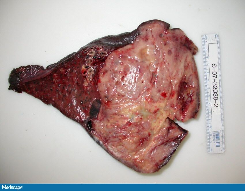

now here are the questions
You are examining a hypoglycemic patient.
CT scanning of the abdomen and pelvis is ultimately performed during
your evaluation; it reveals a mass in the patient's retroperitoneum. If
this mass were to be confirmed as a non-islet cell tumor (NICT), which
of the following findings would NOT be seen in the laboratory
examinations?
Which of the following symptoms would be most likely to occur in the above described patient?
The differential diagnosis in patients who present with syncope is
typically quite broad. Laboratory and other clinical tests, while often
extensive, usually don’t identify the cause. In low-risk patients
without a concerning presentation of syncope, minimal testing is
acceptable and appropriate; however, given this patient’s unusual
presentation and underlying medical conditions, an extensive workup
including evaluation of cardiac, metabolic, and neurologic causes of
syncope was initiated.
A possible
hematoma in the chest wall as suggested by interpretation of the chest
CT scan was initially entertained because this would have explained the
patient’s anemia and possible cardiopulmonary effects (because of the
location); however, this still did not explain the presence of
hypoglycemia. The patient’s anemia was more likely the result of a
chronic gastrointestinal (GI) bleed complicated by anticoagulation. On
cardiac evaluation, his cardiac enzymes, ECG, and echocardiogram did
not reveal any acute processes or conduction abnormalities. Whipple's
triad (hypoglycemic symptoms, low plasma glucose, and relief of
symptoms with the administration of dextrose) was noted. During the
hospital course, his hemoglobulin and hematocrit remained relatively
stable, without any signs of active bleeding after warfarin was
withdrawn.
On review of the patient’s
endocrine data, the presence of a low insulin level was incompatible
with a diagnosis of insulinoma. The plasma insulin and C-peptide levels
were appropriately suppressed in response to the low fasting blood
glucose. His pituitary function was evaluated and revealed a mildly
elevated follicle-stimulating hormone, but normal thyroid hormones,
prolactin, and cortisol levels were noted. Adrenal insufficiency was
also excluded given the combination of a normal morning cortisol level
and normal adrenocorticotropin stimulation test. It was also important
to exclude the possibility of hepatic and renal dysfunction, which can
both lead to recurrent hypoglycemia. Factitious hypoglycemia was not
suspected in this patient as it was excluded by the low levels of
insulin and C-peptide. Additionally, he was not taking any medications
which could have been a source of iatrogenically-induced hypoglycemia.
The
most likely diagnosis was determined to be a non-islet cell tumor
(NICT) producing hypoglycemia. The patient presented with hypoglycemic
symptoms and an abnormal chest examination, which correlated with a
chest mass on CT scanning. Surgical consultation was obtained for a
biopsy of the left-sided chest mass. A CT scan of the abdomen and
pelvis was also obtained to rule out metastatic disease; the result of
the scan was negative. Biopsy of the left chest mass (see Figure 4)
showed bland spindle cells with a lacy or reticulated appearance (see
Figure 5) which led to the diagnosis of a solitary fibrous tumor of
mesenchymal origin. The tumor was noted to be 6.8 × 5.2 × 3.9 in (17.2
× 13.2 × 9.9 cm) in size. This large mesenchymal tumor accounted for
all of the patient’s symptoms, including his recurrent hypoglycemia.
Non-islet cell tumors (NICTs) are a rare but well described etiology of chronic fasting hypoglycemia.
These extrapancreatic tumors are generally of mesenchymal or epithelial
origin. Mesenchymal tumors represent 50% of all cases of NICT. They
include mesotheliomas, fibrosarcomas, rhabdomyosarcomas,
leiomyosarcomas, and hemangiopericytomas. Carcinomas represent another
25% of NICTs and include hepatomas, adrenocortical carcinomas, and
carcinoid tumors. The remaining 25% of NICTs associated with
hypoglycemia include, but are not limited to, hypernephromas, Wilms
tumors, prostate carcinomas, cervical carcinomas, breast carcinomas,
leukemia, lymphomas, and myelomas.
NICTs are characteristically large in size, weighing an average of
4.4-8.8 lb (2-4 kilograms). Over one-third are retroperitoneal in
location, approximately one-third are intra-abdominal, and the
remaining one-third are intrathoracic.
Neuroglycopenic symptoms are the most common clinical features
associated with NICT-induced hypoglycemia. These symptoms include
obtundation, confusion, and behavioral aberrations. The diagnostic study of choice is CT scanning of the suspected tumor location, followed by a tissue biopsy for identification.
In
this patient, the large mesenchymal tumor identified in the
intrathoracic area resulted in the neuroglycopenic symptom of
obtundation. His obtundation was only abated by continuous glucose
administration preoperatively, and by surgical removal of the NICT.
It
has been proposed that NICTs mediate their effects through insulin-like
growth factor (IGF)–II. In normal circumstances, IGF-II is produced by
the liver as a 7.5-kilodalton (kD) molecule. Most IGFs subsequently
form a 150-kD tertiary complex with IGF-binding protein (IGFBP)–3 and
acid-labile glycoprotein. This large complex is retained in circulation
and delivers IGF to tissues, where it interacts with specific IGF
receptors for local growth promotion. Normally, the circulating IGF-II
tertiary complex does not interact with insulin receptors and,
therefore, is not associated with hypoglycemia.
In
contrast to normal physiology, NICTs produce a partially processed,
high molecular weight (MW) IGF-II (also known as “big” IGF-II). Its MW
has been demonstrated to be 11-18 kD. It constitutes up to 50-75% of
circulating IGF-II in patients with NICTs.
Big IGF-II does not form a tertiary complex, but instead forms a binary
complex with less restrictive IGFBPs, such as IGFBP-2. This smaller,
50-kD binary complex allows for capillary crossing and delivery of big
IGF-II to insulin receptors, primarily in skeletal muscle, where
increased bioavailability leads to increased glucose utilization and,
therefore, hypoglycemia. Big IGF-II also binds to insulin receptors in
the liver, where it suppresses gluconeogenesis and glycogenolysis,
thereby enhancing the hypoglycemic response.
The increase in bioavailable IGF-II also leads to the suppression of
insulin and growth hormone, as well as a decrease in the production of
IGF-I, IGFBP-3, and acid labile subunit, while increasing production of
IGFBP-2.
The treatment of hypoglycemia
for patients with NICTs is symptomatic support until resection of the
tumor is performed. During this patient’s hospital course, he required
continuous infusion of dextrose along with frequent monitoring of his
blood glucose. Complete resolution of his hypoglycemic symptoms after
tumor resection supported the diagnosis of NICT-induced hypoglycemia.
Low levels of growth hormone, IGF-I, and IGFBP-3 also supported the
hypothesis that altered IGF-II was the mediator of hypoglycemia.
Although an elevated IGF-II level was not identified, it is well known
that IGF-II levels may be normal or elevated.
In NICT-associated hypoglycemia, IGF-II can cause hypoglycemia at
normal total serum levels as a result of altered processing and
increased bioavailability of IGF-II.
The patient was fortunate to have a benign tumor which did not require further chemotherapy or radiation.
It
should also be noted that this patient’s presentation of hypoglycemia
differs substantially from classic hypoglycemic episodes. Hypoglycemic
symptoms typically include hyperadrenergic and neuroglycopenic-type
symptoms; however, this patient did not describe symptoms such as these
prior to syncope. Patients with tumor-related hypoglycemia usually have
gradual slow falls in their blood glucose. This slow fall does not
trigger the hyperadrenergic response, and neuroglycopenia can progress
from confusion to coma and possible seizures without being recognizable
to the patient. Additionally, it is highly unusual for a patient with
hypoglycemia to spontaneously wake from an altered or comatose state.
Unlike in the setting of hypoglycemia secondary to diabetes
medications, it is likely that in cases of tumor-related hypoglycemia
the body's counter-regulatory mechanisms are able to provide a
sufficient response to bring the serum glucose to a reasonable level
and result in recovery of alertness without pharmacologic intervention.
now here are the questions
You are examining a hypoglycemic patient.
CT scanning of the abdomen and pelvis is ultimately performed during
your evaluation; it reveals a mass in the patient's retroperitoneum. If
this mass were to be confirmed as a non-islet cell tumor (NICT), which
of the following findings would NOT be seen in the laboratory
examinations?
- Decreased insulin level
- Increased C-peptide level
- Normal or elevated insulin-like growth factor (IGF)–II level
- Decreased growth hormone level
Which of the following symptoms would be most likely to occur in the above described patient?
- Altered mental status
- Gastrointestinal pain
- Increased ocular pressure
- Atrial fibrillation
Eman- Admin
- عدد المساهمات : 127
تاريخ التسجيل : 14/09/2009
العمر : 42 -
صفحة 1 من اصل 1
صلاحيات هذا المنتدى:
لاتستطيع الرد على المواضيع في هذا المنتدى